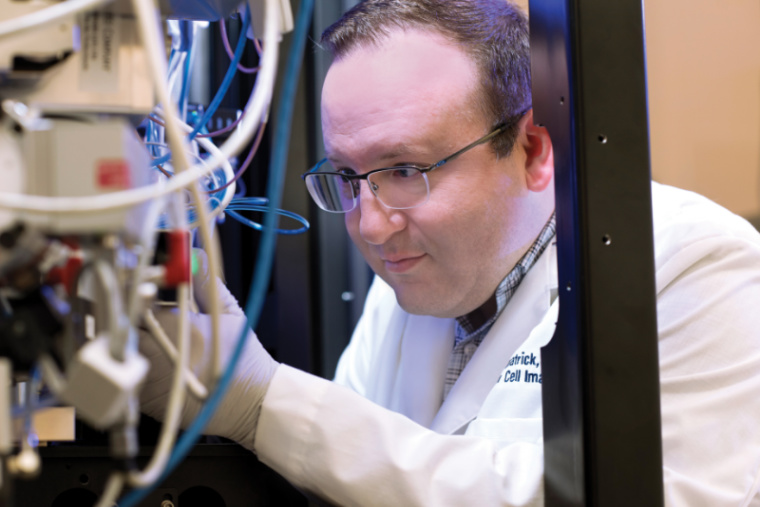
The National Institute of Mental Health has awarded James Fitzpatrick, PhD, a $600,000 grant to purchase a Zeiss LSM 980 Airyscan 2 microscope platform for the Washington University Center for Cellular Imaging (WUCCI). The instrument will complement the existing optical microscopes at WUCCI by expanding super-resolution imaging to a broader range of wavelengths, thereby allowing researchers to use a greater variety of fluorescent probes.
“This is the cutting-edge, newest technology available in the confocal and super-resolution imaging space,” says Fitzpatrick, the scientific director of WUCCI and a professor of neuroscience, cell biology & physiology and biomedical engineering at Washington University School of Medicine in St. Louis. “The grant helps maintain our position as a leading academic imaging center.”
WUCCI provides the full range of imaging capabilities to researchers, from sample preparation to data analysis. The facility houses x-ray, electron, ion and optical microscopes, including the 15-feet-tall Titan Krios cryo-transmission electron microscope, whose custom configuration makes it only one of a handful worldwide.
The LSM 980 Airyscan 2 enables super-resolution microscopy, which surpasses the diffraction limit of light, and provides a resolution down to 120 nm. “If you’re trying to quantify very small structures in the brain, like synaptic morphology, being able to image at higher resolution, beyond the diffraction limit, is important,” says Fitzpatrick.
Other important features of the microscope are the speed at which it can image and the range of wavelengths it is able to detect, in particular, its ability to detect near-infrared fluorescent probes. The new “Multiplex” mode allows for an 8-fold increase in the rate of imaging, which is critically important when imaging highly dynamic processes such as receptor trafficking. In addition, the enhanced near-infrared detection allows for the use of newly developed labels, which are particularly useful for visualizing structures within lipid-dense material, such as the brain and spinal cord.
The new instrument is expected to be installed and ready for use in early 2022.