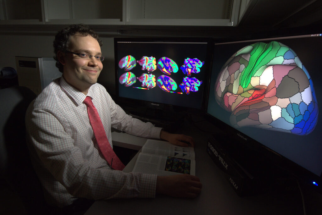
As a graduate student in the Department of Neuroscience, Glasser came to be a leader in the Human Connectome Project.
Matt Glasser was well on his way to starting a career in political science when a flight to South America steered him off course. Glasser was an undergraduate at Emory University at the time and had elected to take a week-long field course in the Amazon rainforest. Wanting a window seat, he switched places with someone else on board and ended up sitting next to an Emory biology professor “whose goal during the flight was to figure out what I wanted to do with my life,” recalls Glasser. Not taking political science for an answer, the professor encouraged Glasser to sign up for a neuroscience course led by a well-known anthropologist, Melvin Konner MD, PhD. Glasser took his advice—“and it was the most interesting class I had in college.”
Glasser scrambled to switch majors and landed up doing a neuroscience honors thesis in the lab of James Rilling, PhD, a professor of anthropology at Emory who studies the evolution of the human brain. Glasser was assigned to analyze data to map the connection between the brain’s language areas, a task that could only be done adjacent to an MRI scanner in the bowels of the university hospital.
With only the company of the scanner, Glasser cemented a new career path, this time in neuroimaging, that led to him becoming an architect of one of the most ambitious endeavors in the history of the field: the Human Connectome Project.
Myelin maps
To pursue his interests in the brain after his undergraduate degree, Glasser decided to go to medical school, but his years as a political science major left him without the pre-requisites to apply. He joined a post-baccalaureate premedical program at Johns Hopkins University to catch up. For a term paper, Glasser related a model from the brain imaging data he’d developed at Emory from connectivity observations to the language deficits from strokes and other lesions. It turned out to yield the worst grade he’d ever received during his academic career—a measly C—because of a difference of opinion with the instructor.
Glasser shrugged it off and published the analysis as part of his first scientific paper, describing the human brain’s language pathways, in 2008 after he returned to Rilling’s group as a lab manager. It remains one of his most popular papers, currently with more than 600 citations according to Google Scholar. “I think a major reason it continues to be cited is because of the material that was in the term paper on brain injury” Glasser says.
This was truly a team effort involving many Human Connectome Project consortium investigators, but I personally credit Matt as being the primary intellectual driver in synthesizing numerous piecemeal advances on diverse fronts into the broader conceptual framework embodied by the HCP-style paradigm.
David Van Essen, PhD
For his graduate work, Glasser knew what he wanted to work on before he even started. At Emory, he had begun investigating how to determine myelin density from MRI scans to help map brain areas. During interviews for medical science training programs (MSTPs), Glasser pitched the idea to David Van Essen, PhD, Alumni Endowed Professor of Neuroscience at Washington University School of Medicine. Van Essen has been a pioneer in charting the terrain of primate brains, first leading the way with exhaustive pencil-and-paper renditions of cortical contours in the 1980s to later advancing the most cutting-edge techniques in imaging. He accepted Glasser’s proposal and Glasser came to WashU in the summer of 2008. “David and I have very similar interests,” Glasser says. “That’s why we have worked so well together.”
Van Essen says the project was a success. “Cortical myelin maps have proven to be extremely useful for identifying many of the cortical areas in our multimodal parcellation and in helping align maps from different individuals onto a common atlas framework,” he says. Glasser has continued to refine the technique, developing a tool to compensate for biases introduced by the MRI scanner. “This new method will likely enable a variety of statistical analyses related to myelin content and how it changes with age and perhaps also in brain disorders,” adds Van Essen.
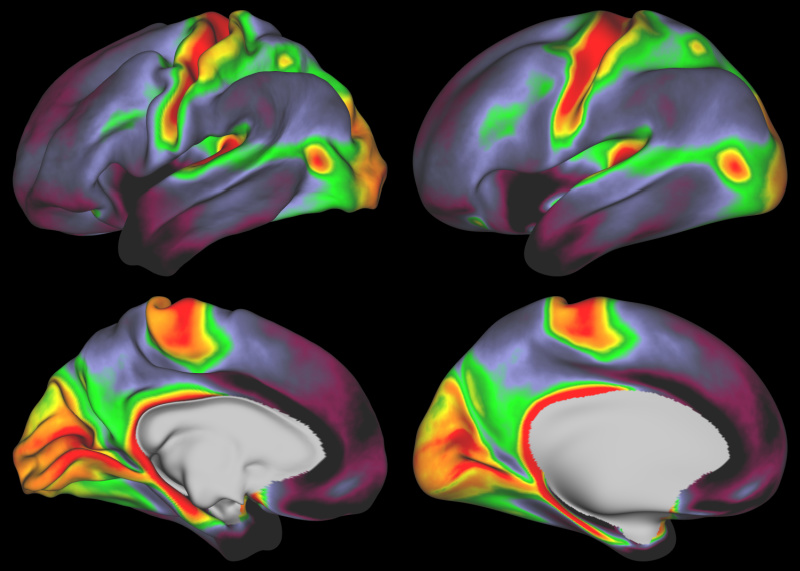
Courtesy Matthew Glasser, MD, PhD and David Van Essen, PhD
The Human Connectome Project
Glasser joined Van Essen’s group at a fortuitous time in its history—in 2009, the National Institutes of Health (NIH) announced that it would be investing $30 million to map the structural and functional connections of the human brain. The timing could not have been more perfect. Glasser and his fellow MSTP students Alex Cohen, MD, PhD, now a neurology assistant professor at Harvard Medical School, and Tim Laumann, MD, PhD, an instructor in the Department of Psychology at Washington University School of Medicine, had been generating connectome data for a competition sponsored by the Organization for Human Brain Mapping (for which they were co-winners), and this effort laid important aspects of the groundwork for WUSTL’s application to the NIH.
In 2010, the agency announced that Washington University, in partnership with the University of Minnesota and the University of Oxford (the so-called WU-Minn-Ox consortium), would lead the Human Connectome Project (HCP)—a grand initiative in which the team would optimize new imaging technologies, generate neuroimaging data from more than 1,000 human subjects and freely share this information with the research community.
The researchers gathered data from different imaging modalities, including task fMRI, resting state fMRI and diffusion MRI. With high-resolution brain scans in hand, Glasser set out to identify the boundaries between different cortical areas, ultimately coming up with 180 areas per hemisphere. The resulting publication graced the cover of Nature in 2016. By the time it appeared in print, Glasser was back to his clinical training and sneaking off to take reporters’ calls about the paper during a medical student rotation in internal medicine. “It was a surreal experience,” he says.
Van Essen says that Glasser was instrumental to developing the HCP-style approach to neuroimaging, which set the standards for acquiring, analyzing and sharing data. “This was truly a team effort involving many HCP consortium investigators,” says Van Essen, “but I personally credit Matt as being the primary intellectual driver in synthesizing numerous piecemeal advances on diverse fronts into the broader conceptual framework embodied by the HCP-style paradigm.” The approach has spread to neuroimaging groups around the world.
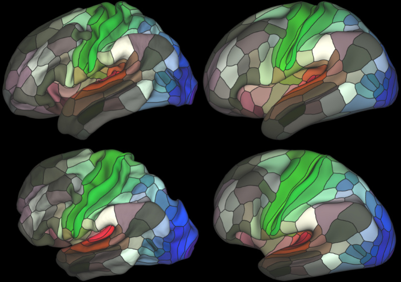
Courtesy Matthew Glasser, MD, PhD, and David Van Essen, PhD
Clinical practice
Although Glasser’s work has had a global influence in the field, St. Louis remains his home. Glasser met his wife, Katherine Holzem MD, PhD, currently senior vascular surgery fellow at BJC, in 2008. They bought a home in 2009 while they were both MSTP students—“It’s a very inexpensive place to live,” he says—within walking distance of the medical school. “It’s great if your commute gives you exercise instead of headaches and traffic.” Their location also places them near some of the best restaurants and cultural activities in the city along Manchester and in the Central West End.
After he completed medical school, he did an internship at St. Luke’s Hospital in Chesterfield—which afforded him several months of research—then returned to Washington University School of Medicine for his residency in radiology. He was selected to participate in the Holman Pathway, a program run by the American Board of Radiology in which residents are afforded up to 21 months of protected research time. It’s the first time the Department of Radiology has had an individual on the Holman Pathway.
Next year, he plans to do a neuroradiology fellowship at WashU as his research continues to focus on using the HCP-style approach to refine the use of myelin maps, removing noise from brain imaging data and understanding variability between people in their cortical areas. He and his wife are joining the WashU faculty in the radiology and surgery departments, respectively. Glasser says that he has stayed at Wash U “because we have a fantastic team here in the lab, a great set of collaborators and the institution has been willing to work with me to make the physician-scientist thing possible.”
Matthew Glasser, MD, PhD
Holman Pathway Resident in Radiology, Washington University School of Medicine
WashU training:
- Medical Science Training Program (MSTP), 2008 – 2017
- Radiology resident 2018 – present
Graduate advisor: David Van Essen, PhD
Expertise: Neuroimaging data analysis methods and neuroanatomy
Favorite St. Louis spot: Rockwell Beer Company
WashU highlight: The Human Connectome Project getting funded