
For decades, neuroscientists considered the visual processing center of the mouse brain simple, even insignificant, compared to that of cats, humans and other animals—and for good reason. The nocturnal animals aren’t known to use their eyes for getting around much, and their brains instead dedicate an enormous amount of real estate to processing information from the animals’ whiskers and ears, while vision is relegated to a tiny shack on the outskirts of town.
It was in these backwaters of the mouse cortex where Quanxin Wang set up shop when he joined the lab of Andreas Burkhalter, PhD, a professor of neuroscience at Washington University School of Medicine, as a postdoctoral researcher in 2001. To trace the routes of neurons in the visual cortex, Wang adopted a technique Burkhalter had developed of flattening the brain’s cortex, much like taking the rind of an orange and slicing it from the edge inward so it can lay flat. “In this way we can see all cortical visual maps in a single tangential section,” says Wang.
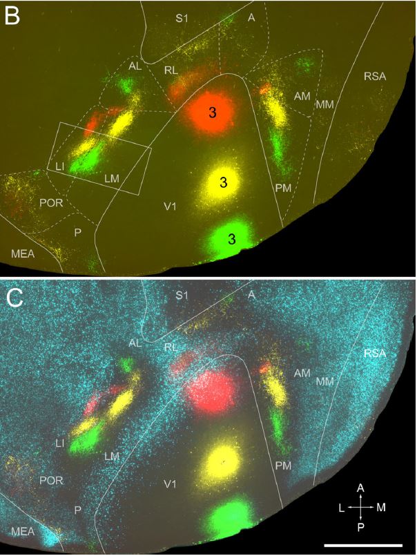
Wang injected three differently colored fluorescent dyes into the primary visual cortex to see where its axonal projections terminated in other cortical areas and found much more complexity than anyone had appreciated. Prior to this work, it was thought that there were two or three extrastriate visual areas in the mouse cortex. Wang’s studies identified at least 10 visual cortical areas, which are hierarchically organized and form dorsal and ventral streams, similar to what’s been observed in primates.
“We basically made the first roadmap of the visual cortex of the mouse,” says Burkhalter. “It then became an overnight sensation because it allowed people to probe parts of the brain they always wanted to.” The paper, published in the Journal of Comparative Neurology in 2007, has been cited several hundred times, according to Google Scholar. Wang’s research in Burkhalter’s lab produced 11 papers.
Now, as a principal scientist at the Allen Institute in Seattle, Wang is continuing to chart the intricate wiring of the mouse brain. Today (October 6) in Nature, he and colleagues published a comprehensive map of the cell types in the upper limb area of the mouse motor cortex, as part of the Brain Initiative Cell Census Network, that includes a wiring diagram of the input and output projections in this area.
“I think where he landed up is really perfect for him,” says Burkhalter. “It gives him the chance to work on something that he is really good at.”
An anatomist at heart
Wang grew up in Heilongjiang Province in China, the son of two physicians. He initially set out to follow in their career footsteps and went to medical school at Heilongjiang University of Traditional Chinese Medicine. Following a master’s degree in acupuncture in the same university, he taught medical students human anatomy for two years as a lecturer in the Department of Anatomy at Harbin Medical University.
Journal of Comparative Neurology, 502(3):339-357, 2007.
Afterward, Wang decided to pursue research, so he left for Japan to earn his PhD at Toyama Medical and Pharmaceutical University. There he joined lab of Osamu Ohtani, PhD, a professor in the department of anatomy, and studied the detailed distribution and ultrastructure of the stomata connecting the pleural cavity and the lymphatics in the rat costal pleura by scanning electron, transmission electron and light microscopy, honing his meticulous approach to anatomical investigations.
He was also introduced to neuroscience by collaborating with the lab of Hisao Nishijo, PhD, a professor in the department of physiology in the same building, on cholinergic and noradrenergic elements in the basolateral nucleus of the rat amygdala. “Once I entered this neuroscience field, I knew the brain is an amazing place and I wanted continue to work in the field,” he says.
After receiving his PhD in 1997, Wang joined the lab of Ichiro Fujita, PhD, a professor at Osaka University, as a postdoc to study visual perception in monkeys. He investigated postnatal development of intrinsic horizontal axons in macaque inferior temporal and primary visual cortices, which are the first and final stages of ventral visual pathways—processing simple and complex stimuli, respectively. He found that intrinsic connections in both visual areas emerge very early on during brain development, even before the animal is born.
During this time, Akiko Yamashita, a friend of Fujita and Wang, was a visiting scientist in Burkhalter’s lab and piqued Wang’s interest in studying in St. Louis. Wang reached out to Burkhalter and packed his bags for what would end up being a decade-long stint at WashU.
Mapmaker of the visual cortex and beyond
Following his work on the layers of the mouse visual cortex, Wang wanted to see where the cells of these layers led. He injected tracers into each visual area to follow their connectivity and identified two structural streams, ventral and dorsal, analogous to what had been observed in the monkey visual system. Again, Wang’s work demonstrated that the mouse brain was more similar to that of primates than was previously known.
Burkhalter says Wang’s success stems from his precise technical skills and the care he took in preparing “absolutely stunning” figures from his fluorescent tracing experiments (the 2007 paper, for instance, made the cover image). “The imagery was so persuasive,” Burkhalter says. “For him, anatomy is its own pursuit.”
Wang went on to contribute to projects in the lab that further revised scientists’ understanding of the mouse visual cortex, first describing the columnar organization of the cortical layers and then demonstrating that visual information is processed in a hierarchical method, with different features successively assembled to ultimately reach the final representation of that object.
Wang says he loved his time at WashU and recalls Burkhalter frequently joining him at the microscope to collect data and train him on techniques. Outside the lab, Wang found St. Louis to be a relaxing city with ample cultural offerings. Among his favorite places is the Missouri Botanical Garden, a 79-acre collection of world-famous gardens and exhibits in the middle of the city. Affordability was a huge plus, too. “For students, they can get a top-notch education, but it costs less to live there than on the East or West Coast. It’s great.”
Cell, 181:936-953, 2020
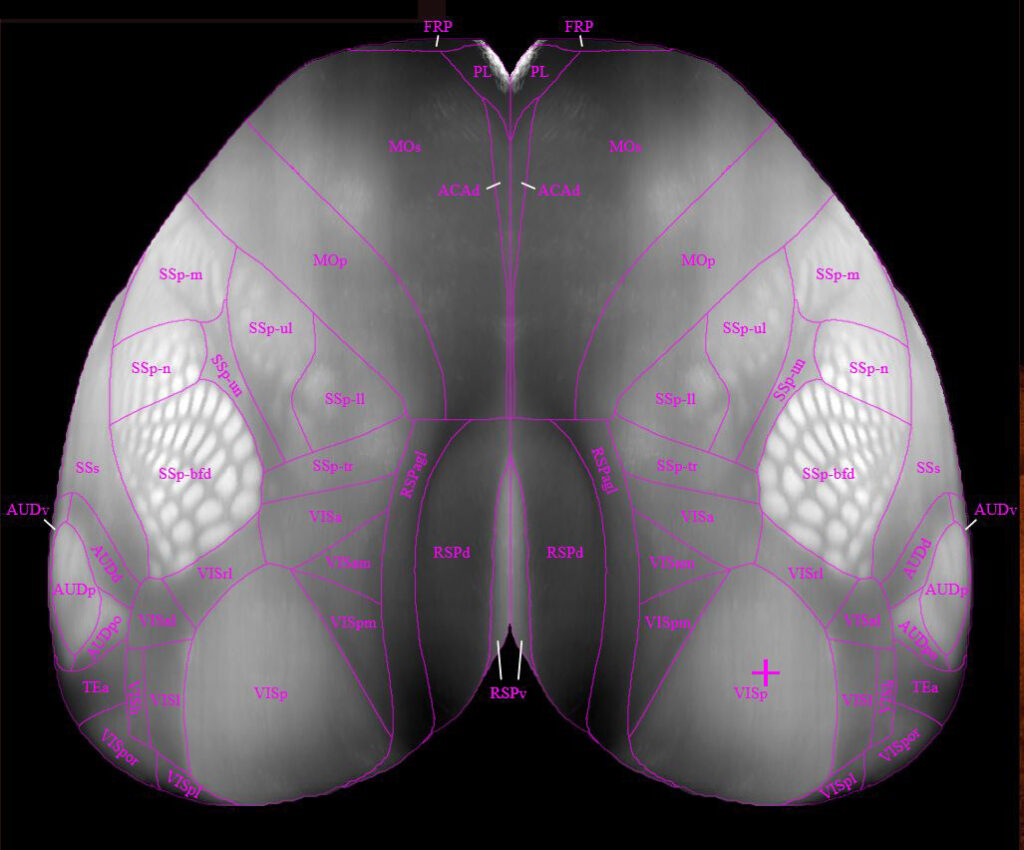
After 10 years at WashU, Wang felt it was time to move on and he joined the Allen Institute in 2010 to begin work on the meso-scale connectome of the mouse brain. He brought with him expertise in neuroanatomy he gained at WashU and shared them with his Allen colleagues to help optimize the tracer injection protocols of the project. Wang has since been part of continued additions to the Allen Mouse Brain Connectivity Atlas and he most recently published a revised 3-D reference map of the mouse brain called the Allen Mouse Brain Common Coordinate Framework version 3 (CCFv3).
In CCFv3, Wang delineated 10 visual cortical areas based on visuotopic maps as he had done in Burkhalter’s lab but in 3D this time. That endeavor involved labeling every structure in each 10-um section of the brain. “The mouse brain is nearly thirteen millimeters long, so that means thirteen-hundred sections we needed to work on,” says Wang. “It’s a lot! It took the team three years to get to the finish line.” He’s now focused on single-cell tracing to identify all the connections each cell type in the mouse brain makes with its targets and using monosynaptic retrograde rabies tracing to investigate whole-brain inputs to a given cell type or class defined by a transgenic line. Wang says his postdoctoral research in Burkhalter’s lab was instrumental in his career progression, and Burkhalter’s support was foundational to his research success at the Allen Institute.
Quanxin Wang, PhD
Principal Scientist, Allen Institute
WashU training: Postdoctoral researcher 2001-2011
Advisor: Andreas Burkhalter, PhD
Expertise: Anatomy of the mouse visual cortex
Favorite St. Louis spot: Missouri Botanical Garden
WashU highlight: Accessibility of faculty