Sunrise to sunset, living organisms use the circadian clock system to modulate their internal daily rhythms. In the fruit fly, Drosophila, a network of 150 dedicated neurons, called pacemakers, must work in perfect synchronization to adapt daily behaviors to changing light conditions. Essential to this process is the neuropeptide PDF, which is released by morning pacemaker neurons to support normal rhythmic behavior. PDF signaling acts in two ways: it synchronizes the molecular clock within pacemakers. Secondly, it delays the neuronal activity of specific subsets of pacemakers away from a Morning to an Evening and a Nighttime activity phase. To do this, PDF signaling needs to persist over periods of many hours and shut down at the right time. It must also adjust this timing each day to reflect seasonal changes in daylight hours. Although its role in the circadian clock is well known, little is understood about what controls these characteristics of PDF signaling.
In a recent paper published in PLoS Genetics, Paul Taghert, PhD, Professor in the Department of Neuroscience at Washington University School of Medicine, and his lab studied the “off” switch for PDF signaling. When they blocked phosphorylation of particular sites on the PDF receptor (PDFR), flies showed altered circadian behavioral patterns, with an increased peak of activity in the morning and delayed evening activity. This discovery shines a spotlight on the importance of identifying what drives circadian signaling events to be shut down.
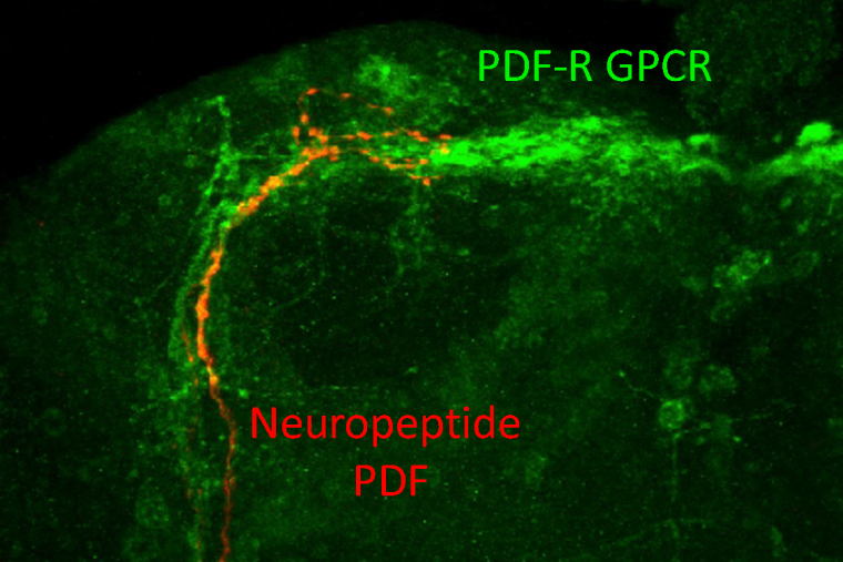
Previous research, including work by Taghert, established how PDF signaling is relayed via PDFR, but questions remained about how PDFR itself is regulated. Taghert and his laboratory sought to illuminate these mechanisms by asking the question, “Each day, how is PDFR signaling turned ‘off?’”
“The on is important, but the off is also,” said Taghert, “because the system has to track the dawn and the dusk to provide the internal timing. The regulation of when PDF signaling starts and stops has to change every day because the sun rises and sets each day at different times.”
Weihua Li, PhD, and Jennifer Trigg, researchers in Tahgert’s lab, approached this question by using a structure-function approach to predict behavioral outcomes. They knew that phosphorylation of GPCRs is well-established as a brake organisms use to reduce downstream signaling, but most studies on neuropeptide receptors have only been conducted primarily in vitro by functional expression in a cell culture model. Using these previous studies as a guide, they sought to identify candidate sites of PDFR receptor de-activation, with the hypothesis that modifying these sites would cause a gain-of-function effect on receptor signaling (i.e., loss of de-activation). To do this, they performed comparative genomic analysis to choose 14 serine, threonine, or tyrosine residues located on the C-terminus that were evolutionarily highly conserved (across fly species that are >60 Myr diverged). They then conducted an “alanine scan”—converting clusters of these residues into alanine to render that locus immune to post-translational modifications including phosphorylation. They created a total of 10 PDFR variants, including simple variants that target one to two clusters and multiple variants, targeting three or more.
The regulation of when PDF signaling starts and stops has to change every day because the sun rises and sets each day at different times.
Paul Taghert, PhD
To test their hypothesis that these variants would cause PDFR gain of function, Li chose to use Drosophila behavior as an in vivo readout of the variants’ effects on circadian rhythms. Either wildtype (WT) PDFR or one of the 10 variants was introduced into a pdfr loss-of-function strain of Drosophila, whose locomotor activity was then tested under three simulated daylight conditions: short day (winter-like), long day (summer-like), and equinox (equal length of light and darkness). Then, to measure the flies’ circadian period without the influence of external light, the flies were subsequently subjected to constant darkness for up to 8 days. The behavioral results supported their predictions—the variant-expressing flies’ activity patterns generally trended in the opposite direction from the behavior of flies lacking pdfr function.
“[It is significant] that we could identify possible phosphorylation sites which, when manipulated, produced anticipated behavioral outcomes based on the previous loss-of-function studies for the whole receptor,” Taghert stated.
Interestingly, the altered behavioral activity rhythms varied by simulated daylight condition and by PDFR variant. In summer-like and winter-like light conditions, flies expressing variants generally displayed greater peaks of activity in the morning and evening, as well as multi-hour delays in their evening activity phase. Under equinox-like conditions, these flies only differed from loss-of-function flies in their morning, but not evening peaks. Once the flies were subjected to constant darkness, many PDFR variants caused 3–4 hour delays to the start of their evening peak with circadian periods 1–2 hours longer than WT controls. This was opposite to pdfr loss-of-function flies, which displayed advanced evening locomotor peaks.
Notably, the location and number of alanine mutations were important. Among the simple variants, alanine mutations closer to the C-terminus caused the most severe phenotypes, while among the multiple variants, the addition of more alanine mutations created stronger effects. These findings suggest that PDFR signaling may be terminated by phosphorylation at specific residues or alternatively by bulk accrual of non-specific phosphorylation, likely in a context-dependent manner. Similar results were recently described for melanopsin, the light sensitive GPCR present in many intrinsically photosensitive retinal ganglion cells of mammals.
Together, these changes to fly activity demonstrate that phosphorylation of PDFR C-terminal residues controls how PDF signaling is curtailed on a daily basis to adapt the circadian clock to changing daylight. The findings provide a predicate for identifying modulatory mechanisms that drive signaling to turn off and emphasize the value of using behavioral outcomes as a readout to identify changes in these signaling events. Future directions include exploring which molecules normally play a role in PDFR modulation and using CRISPR to attach fusion proteins to the receptor to better visualize its activity in vivo. Taghert expressed hope that this research will “start” more studies on how neuropeptide modulation in circadian physiology “stops.”