Skip to content Skip to search Skip to footer
WashU Medicine

Department of Neuroscience

Menu
  • About
    • Vision and Mission
    • Department History
    • St. Louis
    • Contact Us
  • People
    • Faculty
    • Emeritus Faculty
    • Affiliate Faculty
    • Administrative Staff
    • Research Staff
    • Trainees
    • Alumni
  • Research
    • Publications
    • Neuroscience Building
    • Facilities
    • Resources
    • Department and WashU Contacts
  • Education
    • Postdoctoral Research
    • Faculty Awards
    • Graduate Rotations
    • Outreach
    • Body Donor Program
  • Opportunities
    • Positions
    • INSPIRE
  • News & Events
    • Department Events
    • Department News
    • Calendar of Events
  • Giving

Category: Featured publication

Daily activity schedules mapped in the Drosophila brain

January 17, 2023March 6, 2023
Diagram illustrating the functional connections between circadian pacemaker neurons, downstream dopaminergic and neurosecretory centers, and daily behaviors

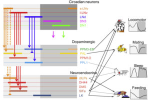
Cellular circuits downstream from circadian pacemaker neurons direct normal daily rhythms of feeding, mating and sleep.

Zebrafish advance as a model organism for Fragile X Syndrome

January 9, 2023March 6, 2023
A zebrafish with its nervous system fluorescently labeled in green and red hunts a paramecium, fluorescently labeled in green

A new study from the Goodhill Lab finds the genetic variant underlying an autism disorder causes changes in fish’s social behavior, preference in visual environment, and neural activity.

Taghert Lab identifies off switch for a set of circadian clock–controlled behaviors in flies

November 30, 2022February 22, 2023
microscope image of the Drosophila brain, fixed and double-stained for PDF (red fluorescence) and PDF-R (green fluorescense)

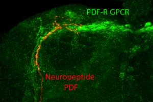
Phosphorylation of a receptor controls how messages from the neuropeptide PDF—which influence dawn and dusk activities in Drosophila—are curtailed on a daily basis to adapt the circadian clock to changing daylight.

People missing all or part of the connection between their brain hemispheres are more persuadable

October 28, 2022February 22, 2023
Brain tractography with connections colored red, green, blue and purple

A study of individuals with congenital corpus callosum dysgenesis suggests they face social difficulties due to being more easily influenced and less aware of being tricked.

Circadian pacemaker neurons exhibit two linked calcium cycles

June 21, 2022July 12, 2022
Schematic of calcium cycles in circadian pacemakers neurons of Drosophila

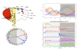
The slow and fast rhythms reflect distinct cellular processes yet nevertheless have a co-phasic relationship.

Study finds key similarities between rodent and human satellite glial cells

May 20, 2022July 12, 2022
Human dorsal root ganglion stained for the neuronal marker TUJ1 (magenta) surrounded by satellite glial cells stained with FASN (green).

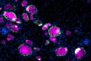
SGCs have been the subject of intense scrutiny for their involvement in inflammation, pain and nerve injury. The results confirm that rodents are a reliable model for translational research on these cells.

Protein linked to intellectual disability has complex role (Links to an external site)

May 17, 2022July 12, 2022
illustration of blue chromosomes on blue background

Finding therapies for fragile X may depend on understanding the many ways the protein’s loss affects the brain.

New primer on logistic models for research in decision neuroscience

March 24, 2022July 12, 2022
Camillo Padoa-Schioppa

Camillo Padoa-Schioppa, PhD, has developed a manual of tools for analysis of economic choices.

Yi lab develops assay to solve mystery genetic variants

March 2, 2022July 12, 2022
Representations of wildtype UBE3A (left) and UBE3A with variant Q588E

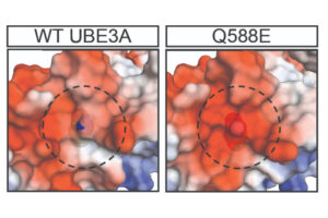
Assistant Professor Jason Yi’s group applied the technique to variants of unknown significance in UBE3A, the gene that underlies Angelman syndrome.

New study shows hierarchical and nonhierarchical network architectures between areas of mouse visual cortex

January 27, 2022February 10, 2022
Rostrocaudal series of coronal sections of the left hemisphere of mouse brain in which the anteromedial area was injected with antergrade tracer BDA.

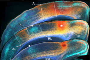
The Burkhalter lab finds that areas are embedded within a hierarchical network in which image fragments from the retina are sent through bottom-up pathways to extract percepts, and top-down pathways from higher to lower areas provide prior knowledge for interpreting the visual scene.

Posts navigation

Previous Page
3 of 5
Next Page

Department of Neuroscience

WashU Medicine

CB 8108

660 S. Euclid Ave.

St. Louis, MO 63110

314-362-3033

Contact Us

  • LinkedIn
  • Twitter

Learn more

Opportunities

Giving

Department Resources

Resources

Directory


©2026 Washington University in St. Louis